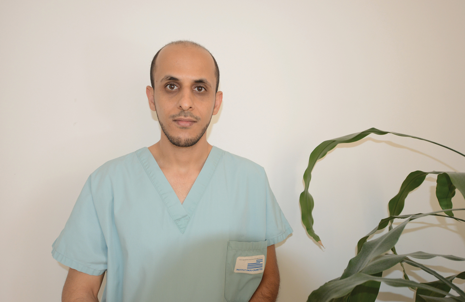

Muscat, Oct 9 - An Omani doctor was part of a team of surgeons that successfully operated on a patient suffering from a rare case of pedunculated giant hepatic hemangioma (tumour). The tumour was discovered in the patient at the Department of Surgical Oncology at the Institute of Paoli Calmettes, in Marseille, France, where Abdullah bin Yahya al Farai is a trainee surgeon. The tumour has been classified as largest of its kind in the world compared with the 19 other registered cases worldwide and published in scientific journals.
Prior to the surgery, the patient was suffering from diarrhoea, vomiting and epigastric pain, which increased while coughing or leaning forward.
The patient had suffered a blunt abdominal trauma at work. Clinical examination revealed a huge abdominal mass occupying most of the epigastric and right hypochondriac region (a part of the abdomen in the upper zone on both sides of the epigastric region and beneath the cartilages of the lower ribs). Blood tests were almost normal. However, the abdominal ultrasound showed a 18 × 10 cm heterogeneous mass in the epigastric region in front of the pancreas and pushing the left liver lobe anteriorly and laterally.
An abdomino-pelvic contrast-enhanced CT scan revealed a prepancreatic tissue tumour with heterogeneous predominantly peripheral enhancement measuring 20 × 9 × 16 cm.
According to Al Farai, the patient had no specific familial and medical personal history, except allergic asthma and active smoking.
However, digestive symptoms persisted and clinical examination confirmed this voluminous palpable abdominal mass.
The case was discussed in the Sarcoma Multidisciplinary Board, and the diagnosis of giant pedunculated liver hemangioma was suspected, and later confirmed. Such benign hepatic tumours are very difficult to diagnose because of their exophytic (tending to grow outward) development.
Al Farai continued, “We decided to perform a liver MRI, which showed several typical hemangioma throughout the liver. The largest one was a pedunculated hemangioma originating from the third hepatic segment, with exophytic development, descending in the epi- and mesogastric region, pushing the pancreas backwards. The dimensions were 20 × 8 × 14 cm, the largest among reported cases worldwide.” Due to the risk of torsion (twisting) and rupture, surgical excision was performed without any preoperative biopsy. Subsequently, via a large median laparotomy incision, a left hepatic lobectomy (surgical removal of a lobe of an organ) was carried out.
The patient was discharged from the hospital on the fourth day after the surgery. A team of three surgeons participated in the surgery.
Al Farai said, “It was an exceptional experience, being part of the surgical team responsible for handling a rare case. The case my team has successfully treated was the largest among them, which makes it a reference case worldwide.” Dr Al Farai is a graduate of Sultan Qaboos University and is about to complete his General/Internal Surgery specialisation at the Aix-Marseille University, France.
He is also preparing for a specialisation in liver and pancreatic surgery at the Department of Surgical Oncology at the Institut Paoli-Calmettes.
Oman Observer is now on the WhatsApp channel. Click here


